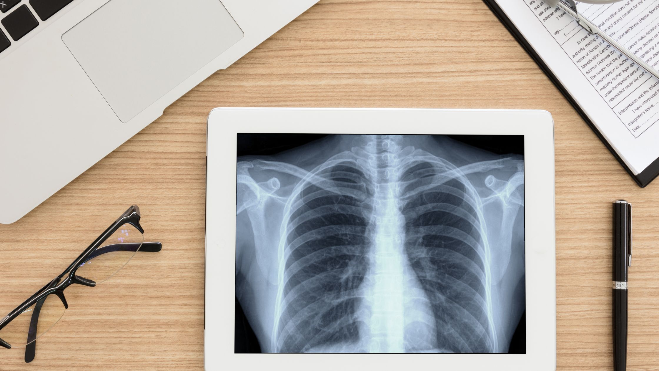
X-rays are a form of electromagnetic radiation, similar to visible light. Unlike light, however, x-rays have higher energy and can pass through most objects, including the body. Medical x-rays are used to generate images of tissues and structures inside the body. If x-rays traveling through the body also pass through an x-ray detector on the other side of the patient, an image will be formed that represents the “shadows” formed by the objects inside of the body.
One type of x-ray detector is photographic film, but there are many other types of detectors that are used to produce digital images. The x-ray images that result from this process are called radiographs.
How do medical x-rays work?
To create a radiograph, a patient is positioned so that the part of the body being imaged is located between an x-ray source and an x-ray detector. When the machine is turned on, x-rays travel through the body and are absorbed in different amounts by different tissues, depending on the radiological density of the tissues they pass through.
Radiological density is determined by both the density and the atomic number (the number of protons in an atom’s nucleus) of the material being imaged.
For example, our bones contain calcium, which has a higher atomic number than most other tissues. Because of this property, bones readily absorb x-rays and therefore produce high contrast on the x-ray detector. As a result, bony structures appear whiter than other tissues against the black background of a radiograph. Conversely, x-rays travel more easily through less radiologically dense tissues, such as fat, muscle, and air-filled cavities such as the lungs. These structures are displayed in shades of gray on a radiograph.
When are medical x-rays used?
Listed below are examples of examinations and procedures that use x-ray technology to either diagnose or treat disease:
-
Diagnostic
X-ray radiography: Detects bone fractures, certain tumors and other abnormal masses, pneumonia, some types of injuries, calcifications, foreign objects, or dental problems.
Mammography: A radiograph of the breast that is used for cancer detection and diagnosis. Tumors tend to appear as regular or irregular-shaped masses that are somewhat brighter than the background on the radiograph (i.e., whiter on a black background or blacker on a white background). Mammograms can also detect tiny bits of calcium, called microcalcifications, which show up as very bright specks on a mammogram. While usually benign, specific patterns of microcalcifications could indicate the presence of cancer. Learn more about mammography here.
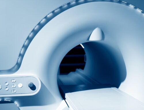
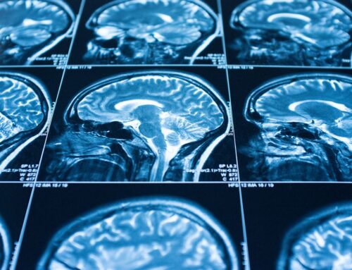
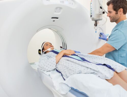
Computed tomography (CT): Combines traditional x-ray technology with computer processing to generate a series of cross-sectional images of the body that can later be combined to form a three-dimensional x-ray image. CT images are more detailed than plain radiographs and give doctors the ability to view structures within the body from many different angles. Learn more about CT here.
Fluoroscopy: Uses x-rays and a fluorescent screen to obtain real-time images of movement within the body or to view diagnostic processes, such as following the path of an injected or swallowed contrast agent. This technology is also used with a radiographic contrast agent to guide an internally threaded catheter during cardiac angioplasty, which is a minimally invasive procedure for opening clogged arteries that supply blood to the heart.
-
Therapeutic
Radiation therapy in cancer treatment: X-rays and other types of high-energy radiation can be used to destroy cancerous tumors and cells by damaging their DNA. The radiation dose used for treating cancer is much higher than the radiation dose used for diagnostic imaging. Therapeutic radiation can come from a machine outside of the body or from a radioactive material that is placed in the body, inside or near tumor cells, or injected into the blood stream.
How do medical x-rays work?
To create a radiograph, a patient is positioned so that the part of the body being imaged is located between an x-ray source and an x-ray detector. When the machine is turned on, x-rays travel through the body and are absorbed in different amounts by different tissues, depending on the radiological density of the tissues they pass through.